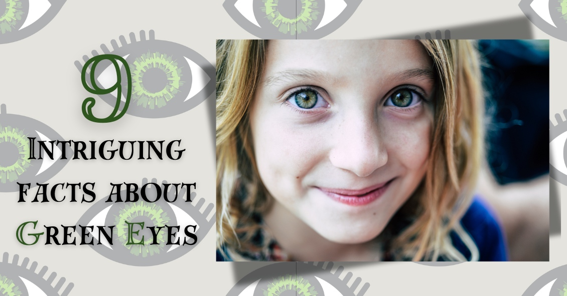
In honor of St. Patrick’s Day and the “wearin’ of the green,” we thought it would be fun and fitting to share some interesting facts about green…eyes!
#1
Green is the rarest eye color. If your sparklers are truly green, you are something of a unicorn… only about 2% of the world’s population sport this hue.
#2
Green-eyed people can be found all around the globe. There is a Chinese village, Liqian, where a high percentage of the population sports green eyes and lighter hair.
#3
There isn’t any actual green pigment in a green eye. Melanin, a natural pigment that helps determine our skin, hair, and eye color, is found in all eyes. Brown eyes have quite a lot and blue eyes have relatively little. Green eyes are also low on melanin, but in addition they contain lipochrome, a yellowish, fat-soluble pigment. Lipochrome is also found in things like butter, eggs, and corn. So a little melanin, some lipochrome, and a cool light dispersing scattering called the Tyndall Effect combine to produce those rare green eyes!
#4
At least 16 genes contribute to eye color. You might have been taught in biology class that two brown-eyed parents can have only brown-eyed children, but it’s more complicated than that.
#5
Green eyes are popular in cultural references. Here are some famous characters with green eyes:
· Jane Eyre—that plucky governess living in a “haunted” mansion, from the book Jane Eyre
· Rapunzel—another courageous hero in Tangled
· Scarlet O’Hara—feisty protagonist in Gone with the Wind
· Scar—the scheming uncle in The Lion King
· Mary Jane Watson, Catwoman, Batgirl—green eyes are popular in the world of comics
· Sara Crewe—brave little girl from A Little Princess
· Harry Potter – from the Harry Potter book series by J.K. Rowling
#6
In a large survey performed by All About Vision, green was voted the most attractive eye color, with over 20% of the 66,000 respondents choosing this hue.
#7
More women than men have green eyes. Scientists aren’t sure why this is, but it suggests there is an underlying gender-related factor that causes this difference.
#8
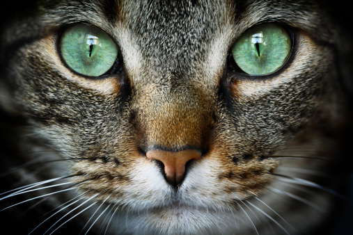 Animals can also sport green eyes. Green eyes occur in dogs, snakes, frogs, birds, monkeys, multiple members of the cat family, and many other animals.
#9
People with green eyes are more likely to have certain health issues. Green eyes are more prone to melanoma of the uvea, a type of eye cancer, than are dark eyes. The same is true for macular degeneration. If you have green eyes, protect those beautiful peepers with a quality pair of sunglasses to lower your chances for these diseases!